
COMPREHENSIVE VESTIBULAR SYSTEM TESTING
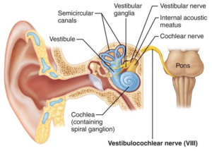
Head injuries and injuries to the neck (WAD I or II) can lead to disruption of the visual and auditory systems. The Vestibular System is a sensory system that consists of 3 canals of the inner ear plus otoliths (saccule and utricle) connected to the visual system. It is responsible for maintaining a sense of position and balance by providing the brain with information about motion, head position and spatial orientation.
The most common complaint post-concussively is “dizziness”. Dizziness is often used to describe multiple symptoms including vertigo (the illusion of movement), disequilibrium (unsteadiness or imbalance) and lightheadedness (pre-syncope). Symptoms can last for 6 months or more and the cause can be elusive unless all parts of the vestibular system.
Other symptoms include Nystagmus (involuntary movement of the eyes), Motion Sickness and Hearing loss.
Vestibular function testing is invaluable at arriving at a proper diagnosis and ultimately the optimal treatment for affected individuals.
(Several good articles can be found here.)
Anatomy of the Vestibular System
Semicircular Canal System – 3 canals, in orthogonal planes, responsible for coordinating rotational movement.
Otoliths – aka “ear stones” are located in the Vestibule and detect linear acceleration.
Vestibulocochlear Nerve: aka the eighth cranial nerve transmits sound and equilibrium information to the brain.
The information is passed through vestibulospinal system in the spinal cord to maintain posture and balance.
Testing Balance and Verticality
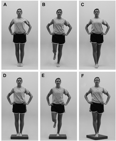
Balance Error Scoring System and Posturography are two tests designed to assess the effects of concussion on static postural stability. The testing consists of 6 tests of standing balance on both a firm and a foam surface.
A final test, Subjective Visual Vertical, consists of looking at a vertical straight line and deciding what is purely vertical and what isn’t. This tests the function of the utricle (one of the two otolith organs) which is responsible for providing a sense of directionality.
Testing the Vestibulo-Ocular Reflex (VOR)
Eyes respond to head movement reflexively. If the head moves left the eyes move right automatically to maintain focus on an object. This is known as the Vestibulo-Ocular Reflex (VOR) and can be impaired following a head injury. Generally one eye is found to lag behind the other.
A Suppression Head Impulse Test:
The investigator elicits small but unpredictable head movements from the patient to stimulate the VOR. A dysfunction is the result of a semicircular canal injury.
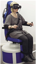
Step Rotation Chair:
Patient is seated in a rotating chair in a darkened room. The rotation triggers a nystagmus (rhythmic and involuntary eye movement). The time take to recover is a a measure of the health of the VOR and indicates an issue with either the brain or the inner ear.
Oculo-Motor Testing
The visual and vestibular systems are intimately connected. Specific eye movements are examined to further identify the cause of problems.
Saccadic Testing:
Rapid eye movement between two stable positions, such as moving from one word to the next when reading text.
Smooth Pursuit:
Allowing the eyes to closely follow a moving object. Injury to the cerebellum will produce a nystagmus.
Optokinetic Testing:
Combination of successive Pursuits and Saccades. Assesses the patients ability / inability to move the eyes in a normal manner under a variety of circumstances.